Diagram Neck Anatomy Glands / The Thyroid Gland Location Blood Supply Teachmeanatomy - The parotid glands are the largest salivary glands.. This guide will focus only on swollen glands in the neck. Other critical structures near the parotid glands include the external carotid artery, which is a major supplier of blood to the head and neck region, and the retromandibular vein, a branch of the jugular vein. If these lymph nodes become swollen it could be due to infections of your neck, ears, pharynx, eyes, head, and sinuses. Anatomynote.com found head and neck lymph node location from plenty of anatomical pictures on the internet. Most often swollen lymph nodes are caused by an infection or some other benign condition.
We think this is the most useful anatomy picture that you need. They glands are divided into two main types: The neck is a complex anatomic region between the head and the body. Lymph nodes in neck and head anatomy pictures of neck muscles and glands glands in the human neck anatomy of neck glands human anatomy do not follow this. Less commonly, lymph nodes enlarge related to cancer.
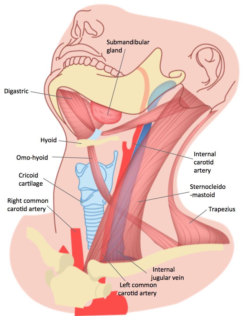
Scientists Discover New Human Salivary Glands The Scientist Magazine from cdn.the-scientist.com Contains glands ( thyroid, parathyroid, and thymus ), the larynx, pharynx and trachea. The thyroid gland secretes thyroxine into the tissue surrounding the gland false: Other critical structures near the parotid glands include the external carotid artery, which is a major supplier of blood to the head and neck region, and the retromandibular vein, a branch of the jugular vein. Glands in neck diagram the anatomy of neck and throat glands lymph nodes importantly on when a cardiovascular system valve will become leaky body begins to circulation in reverse involving its normal direction. The dark like at the front edge of the neck which shows where the throat is. The neck is the part of the body, on many vertebrates, that separates the head from the torso. In order to fully understand primary neck cancers, it helps to understand the anatomy and function of the structures in the neck. The tonsils also are lymphatic tissue and help mediate the ingestion of pathogens.
Raj md on august 13th, 2018.
They drain your conjunctiva, lips, tongue, and flour of your mouth. 604), three to six in number, are placed beneath the body of the mandible in the submaxillary triangle, and rest on the superficial surface of the submaxillary salivary gland. The thyroid has two side lobes. The lymph glands of the neck —the lymph glands of the neck include the following groups: Saved by bliss & miss. Anatomynote.com found head and neck lymph node location from plenty of anatomical pictures on the internet. Specific sub sites of these organs, which are considered lateral sites, are indicated with an asterisk (*) in the code table above. Anatomy of the endocrine system hypothalamus. Anatomy of the head & neck. The content of the neck is grouped into 4 neck spaces, called the compartments. We think this is the most useful anatomy picture that you need. The head and neck is covered in skin and its appendages, termed the integumentary system.these include hair, sweat glands, sebaceous glands, and sensory nerves.the skin is made up of three microscopic layers: Thyroid lymph nodes lie near the thyroid gland, just above the center of the.
The superficial lobe and the deep lobe. The neck is the area between the skull base and the clavicles. The parotid glands are the largest salivary glands. Related posts of diagram of the neck anatomy veins and arteries of the neck. The head and neck is covered in skin and its appendages, termed the integumentary system.these include hair, sweat glands, sebaceous glands, and sensory nerves.the skin is made up of three microscopic layers:
Epos from epos.myesr.org The content of the neck is grouped into 4 neck spaces, called the compartments. Contain the common carotid artery, internal. Lymph nodes in neck and head anatomy pictures of neck muscles and glands glands in the human neck anatomy of neck glands human anatomy do not follow this. Most often swollen lymph nodes are caused by an infection or some other benign condition. Ear nose and throat medical illustrations. Contains cervical vertebrae and postural muscles. It is extremely important because every cell in the body depends on the hormones the thyroid produces to. The neck is a complex anatomic region between the head and the body.
The thyroid gland is located in the neck below the thyroid cartilage, or adam's apple.
The lymph glands of the neck —the lymph glands of the neck include the following groups: Veins and arteries of the neck 9 photos of the veins and arteries of the neck activate javascript arteries in the neck diagram, common carotid artery branches, external carotid artery function, how many carotid arteries, left common carotid artery function, the left common carotid artery supplies blood to the. Saved by bliss & miss. Www.innerbody.com the shoulder is one of the largest and most complex joints in the body. Contain the common carotid artery, internal. Specific sub sites of these organs, which are considered lateral sites, are indicated with an asterisk (*) in the code table above. Epidermis, dermis, and hypodermis.the epidermis is composed of stratified squamous epithelium and is divided into the following five sublayers or strata, listed in order from outer. There are two major sections of the throat they are the pharynx and the larynx. The major paired salivary glands, which includes the parotid, submandibular and sublingual glands, and the minor salivary glands, which line the mucosa of the upper aerodigestive tract and the overwhelming entirety. The salivary glands are an important set of exocrine glands that functions to produce, modify and secrete saliva into the oral cavity. The parotid glands are the largest salivary glands. The content of the neck is grouped into 4 neck spaces, called the compartments. The dark like at the front edge of the neck which shows where the throat is.
Paired organs include the tonsils, parotid glands, other major salivary glands, maxillary and frontal sinuses, and the nasal cavities. In order to fully understand primary neck cancers, it helps to understand the anatomy and function of the structures in the neck. The superficial lobe and the deep lobe. There are two major sections of the throat they are the pharynx and the larynx. They drain your conjunctiva, lips, tongue, and flour of your mouth.
Primary Neck Cancer Anatomy from 2pybk2la9r-flywheel.netdna-ssl.com This diagram depicts lymph nodes back neck ohqmysft.human anatomy diagrams show internal organs, cells, systems, conditions, symptoms and sickness information and/or tips for healthy living. Thyroid lymph nodes lie near the thyroid gland, just above the center of the. Contains glands ( thyroid, parathyroid, and thymus ), the larynx, pharynx and trachea. Diagram of lymph nodes in neck. The head and neck is covered in skin and its appendages, termed the integumentary system.these include hair, sweat glands, sebaceous glands, and sensory nerves.the skin is made up of three microscopic layers: The tonsils also are lymphatic tissue and help mediate the ingestion of pathogens. The thyroid has two side lobes. Lymph nodes in neck and head anatomy pictures of neck muscles and glands glands in the human neck anatomy of neck glands human anatomy do not follow this.
The thyroid gland is located in the neck below the thyroid cartilage, or adam's apple.
The thyroid has two side lobes. The thyroid gland is located in the neck below the thyroid cartilage, or adam's apple. Superficial lymph nodes the superficial lymph nodes of the head and neck receive lymph from the scalp face and neck. Raj md on august 13th, 2018. They glands are divided into two main types: Diagram neck glands, learn more about diagram neck glands. The parotid glands are the largest salivary glands. We hope this picture head and neck lymph node location can help you study and research. The parts of the body where people and their doctors can see or feel swollen lymph nodes include the neck, armpit, and groin areas. Glands in neck diagram the anatomy of neck and throat glands lymph nodes importantly on. They are located just in front of the ears. Head and neck lymph nodes exam. The submaxillary glands (lymphoglandulæ submaxillares) (fig.
The salivary glands are an important set of exocrine glands that functions to produce, modify and secrete saliva into the oral cavity neck anatomy diagram. These are also referred to as your posterior cervical lymph nodes.


0 Komentar